English
 German
 Spanish
 Italian
 Japanese
 United states
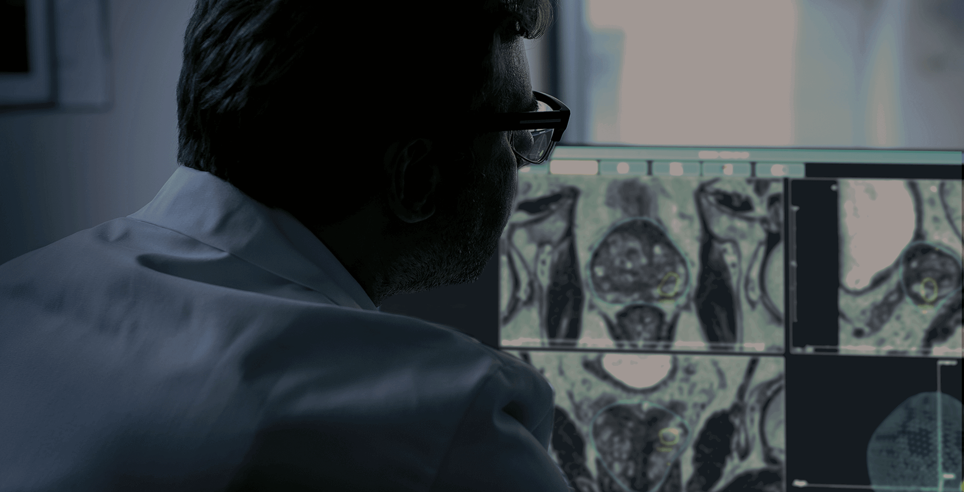
According to the National Cancer Institute, a fusion biopsy procedure combines pictures from an MRI scan and an ultrasound to create a detailed view of the prostate. This procedure aims to make it easier to identify abnormal tissue and guide the biopsy needle into the specific area. Fusion biopsy may help detect prostate cancer at an early stage and assist in planning treatment.
Today, there are two types of fusion biopsy: cognitive or software–assisted fusion. In this article, we will explore the topic of fusion biopsy in more detail and all that it involves.
Differences between cognitive fusion and software assisted fusion
Identifying lesions directly on an MRI image, and then using the ultrasound image to select biopsy samples requires skills from the physicians to mentally superimpose the two images. This process is known as cognitive fusion. While this technique can be quite effective for an experienced urologist, it may prove challenging for a less experienced one, and there is a risk of error. Since both the patient and the prostate can move, compensating for these movements is essential to maintain accuracy. With cognitive fusion, this is nearly impossible, as there is no real-time update of the prostate’s position.
This is why most urologists use software fusion, where MRI and ultrasound images are automatically superimposed to provide physicians with the clearest possible view of the prostate. There are two types of software fusion: elastic and rigid fusion. Rigid fusion superimposes the ultrasound and MRI images without adjusting them to fit each other. Elastic fusion, on the other hand, aligns images automatically, providing urologists with a precise map of the prostate. With Koelis Trinity®, and our OBT (Organ-Based Tracking) Fusion® technology, this 3D map is updated with each sample. During every prostate biopsy, the technology automatically compensates for patient movements and prostate deformations, recording the exact location of each core with millimetric accuracy.
“I have used both and I just think elastic fusion is more accurate. If you know you're hitting the target, you take fewer biopsies, that's better for the patient because it's less uncomfortable.”
Dr Simon Bott, Frimley Park Hospital, Surrey (United Kingdom)1
The benefits of elastic fusion
It has been proven that elastic fusion increases the accuracy of prostate biopsy and cancer detection2.
The study led by Dr. Marco Oderda (Italy) between 2010 and 2017 showed that using elastic fusion results in effective cancer detection, and incorporating systematic cores improves the cancer detection rate by 13% for all cancer and 9% for significant prostate cancer3.
Elastic fusion also improves patient comfort during the procedure. By providing a precise up-to-date map of the prostate, it increases accuracy and reduces the number of cores needed to cover the lesion. As a result, the procedure is shorter and less painful for the patient.
Additionally, collecting fewer cores reduces the pathologist’s workload, fostering better collaboration. This is particularly valuable in the UK, where access to pathologists is limited1.
With our patented OBT Fusion® technology, our software fusion enhances accuracy and provides real-time tracking of both the lesion and the needle. Automatically performed during each prostate biopsy, it compensates for patient movements and prostate deformations, recording each core location with millimetric precision.
Using fusion biopsy for offering personalized care
Obtaining a precise image of the prostate and biopsy cores enables physicians to offer personalized care to their patients. The number of biopsies depends on the size and the stage of the lesion. For example, in a 50-year-old man undergoing a prostatectomy or a radical treatment, the physician may take more biopsies around the neurovascular bundles. For a patient who is candidate for brachytherapy, doctors might focus on taking more biopsies around the urethra. In the case of an elderly gentleman with a T3A tumor, possibly located anteriorly, physicians can take just two or three cores, confident they have targeted the lesion without needing additional biopsies. This approach clearly reduces the patient discomfort, shortens the physician’s operative time, and minimizes the pathologist’s workload1.
To conclude, fusion biopsy is quite a standard in prostate biopsy, but elastic is more efficient than cognitive and rigid fusion. It is now proven, and it is time to adopt it!
Sources :
- 1 Podcast Dr Bott /https://podcast.ausha.co/prostate-talk/unveiling-the-power-of-fusion-biopsies
- 2 https://pubmed.ncbi.nlm.nih.gov/36259316/
- 3 Accuracy of elastic fusion biopsy in daily practice: Results of a multicenter study of 2115 patients – Oderda et al., Int J Urol, August 2018