
 English
 German
 Spanish
 Italian
 Japanese
 United states
As the field of prostate cancer diagnostics continues to evolve, healthcare providers are under increasing pressure to improve procedural efficiency while maintaining a high level of clinical precision. MRI-ultrasound fusion procedures rely heavily on accurate prostate contouring and segmentation, making workflow consistency and image fusion quality essential components of successful biopsy and focal therapy procedures.
To help address these challenges, ProMap Smart introduces a new approach to automated prostate contouring powered by artificial intelligence (AI). Designed to easily integrate with the KOELIS Trinity® system, ProMap Smart leverages AI-assisted automation to streamline prostate segmentation and support more efficient MRI-US fusion procedures. Continue reading to learn more about ProMap Smart and its revolutionary prostate contouring capabilities.
What is ProMap® Smart?
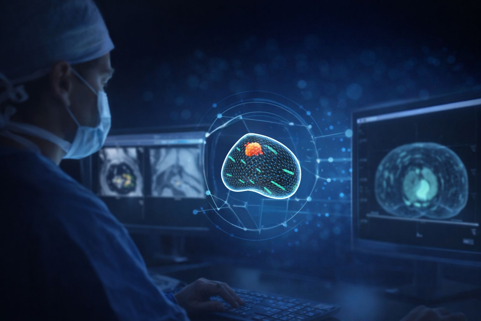
ProMap Smart is designed to advance MRI-ultrasound fusion procedures through AI-assisted prostate contouring. Integrated directly within the KOELIS Trinity platform, ProMap Smart automates one of the most critical and time-intensive steps in prostate fusion procedures to help improve workflow efficiency and procedural confidence.
Built on a patented hybrid engine combining Machine Learning and 3D Elastic Modeling, ProMap Smart is trained on more than 8,000 annotated prostate MRI datasets from expert medical centers worldwide¹. The software achieves 98% accurate prostate segmentation in under five seconds², helping physicians streamline MRI-US fusion workflows while supporting precise needle guidance during prostate biopsy and focal therapy procedures.
ProMap Smart introduces a new level of automation to prostate fusion procedures by reducing the need for time-intensive manual contouring and helping streamline the MRI-US fusion workflow. Its combination of speed, workflow integration, and procedural consistency positions ProMap Smart as an important advancement in the evolution of prostate cancer diagnostics and image-guided intervention.
Benefits of AI in Prostate Cancer Workflows
Artificial intelligence is helping improve prostate cancer workflows by automating complex imaging tasks that traditionally required significant physician time and experience. By reducing manual workflow steps, AI-assisted technologies can help increase efficiency and support more standardized imaging and fusion procedures across clinical teams.
AI also has the potential to enhance procedural confidence by supporting more reproducible prostate segmentation and contouring. As these technologies continue to evolve, they are becoming an increasingly important part of modern prostate cancer diagnostics, biopsy guidance, and treatment planning.
The Importance of Workflow Efficiency
Workflow efficiency plays a critical role in modern prostate cancer diagnostics, particularly as healthcare providers manage increasing patient volumes and growing demand for MRI-guided procedures. Time-intensive manual steps during prostate fusion workflows can introduce variability, increase procedure time, and place additional demands on clinical staff.
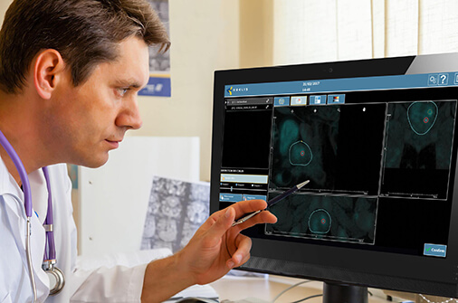
In MRI-ultrasound fusion procedures, efficient prostate segmentation and contouring are essential for maintaining procedural flow while supporting accurate image fusion and biopsy targeting. AI-assisted technologies are helping streamline these workflows by reducing manual processing steps and improving consistency across procedures.
Prostate Contouring vs. Segmentation
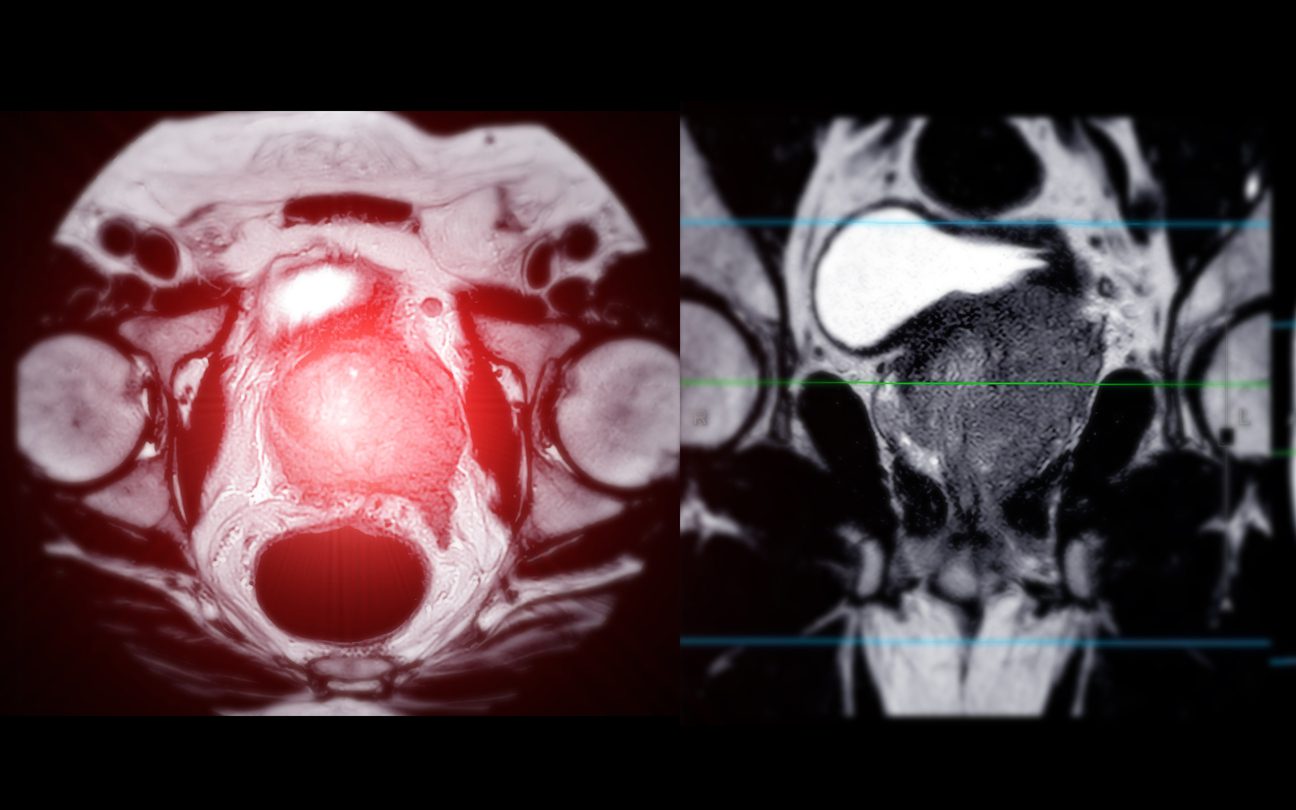
In prostate fusion procedures, the terms prostate segmentation and prostate contouring are often used together, but they describe slightly different parts of the imaging workflow. Prostate segmentation refers to the process of separating the prostate gland and allowing the system to recognize the boundaries of the organ in three dimensions. This step forms the foundation for accurate MRI-ultrasound fusion, 3D modeling and needle guidance during prostate biopsy and focal therapy.
On the other hand, prostate contouring refers more specifically to the outlining or tracing of the prostate boundaries on imaging slices. Traditionally, this process is performed manually by the physician and can vary depending on user experience and workflow preferences.
AI-assisted technologies such as ProMap Smart help automate this process, thereby reducing manual workflow burden while supporting more consistent and reproducible fusion procedures within the KOELIS Trinity system.
AI in Prostate MRI
Artificial intelligence is playing an increasingly important role in prostate MRI by helping clinicians analyze imaging data more efficiently and consistently. As prostate MRI has become a critical component of modern prostate cancer diagnosis and treatment planning, AI-assisted software is being developed to support tasks such as prostate segmentation and lesion detection.
One of the most promising applications of AI in prostate MRI is its ability to assist with MRI-ultrasound fusion procedures used during targeted prostate biopsy and focal therapy. Accurate prostate segmentation and contouring are essential for reliable MRI-US fusion, making AI-assisted automation valuable for supporting procedural consistency and reducing manual workflow burden.
Technologies such as ProMap Smart represent part of this broader shift toward integrating AI directly into image-guided prostate cancer workflows where the focus is not only on speed, but also on improving precision and clinical confidence.
References:
¹ Evaluation of the performance of ultrasound and MRI AI models (ProMap® SMART), conducted on 386 independent test cases annotated by expert clinicians, 2025.
² Dataset size information extracted from the internal Koelis Prostate Contour Inference data report, 2025.