English
 German
 Spanish
 Italian
 Japanese
 United states
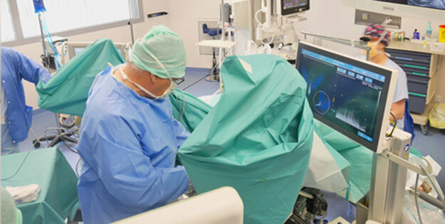
Prostate cancer screening and treatment has come a long way with modern medicine and innovative technology. In fact, the first prostate biopsy on record took place in the early 20th century using the transperineal approach¹ (known as the open perineal prostate biopsy), and the transperineal biopsy was long-considered the gold standard.
However, the early transperineal approach came with risks and drawbacks since it required an incision on the perineum, causing patient discomfort and pain during and after biopsy. Thus, technical modifications and advancements in ultrasound technology led to the development of the transrectal ultrasound biopsy (TRUS), which took the place as the gold standard biopsy method.
But now, innovative medical device technology contributes to the resurgence of the transperineal biopsy with MRI-ultrasound guidance. This advanced method of prostate cancer biopsy uses MRI fusion to fuse the ultrasound and MR image sets together for a better visual representation of the prostate and fewer patient complications.
Are you curious about the transperineal biopsy, how it works, and how it compares to the transrectal approach? Keep reading to find out answers to these questions and more.
What is a Transperineal Prostate Biopsy?
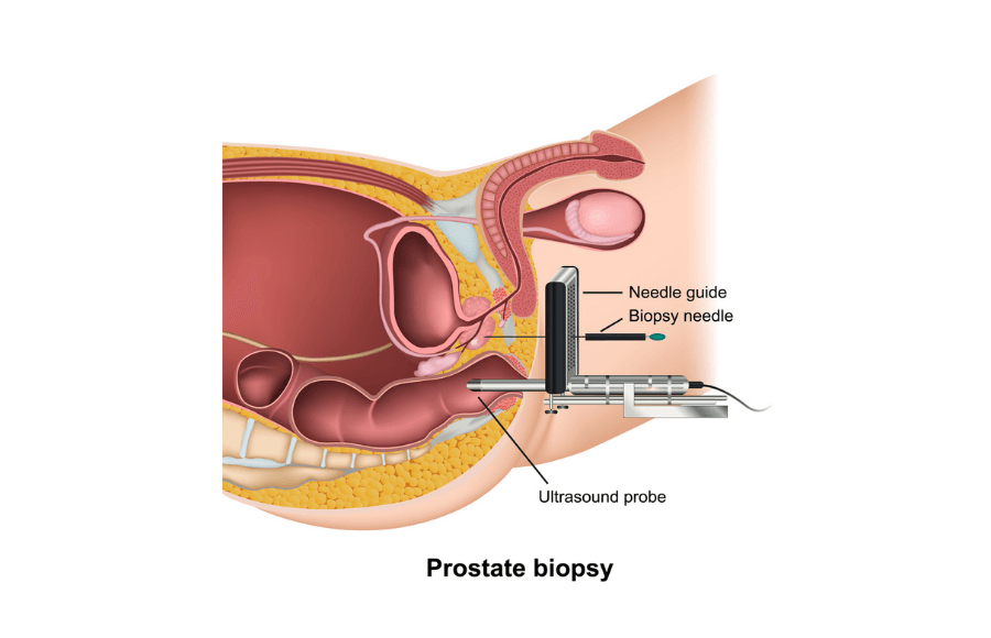
Above: Example diagram of a transperineal prostate biopsy. Notice the needle sampling the prostate by entering through the perineum with the ultrasound probe in rectum.
A transperineal prostate biopsy is a method of screening for prostate cancer that’s become increasingly popular among urologists. While the majority of prostate cancer biopsies are still performed transrectally, the transperineal approach is innovative and better suited for patients to minimize infection and discomfort.
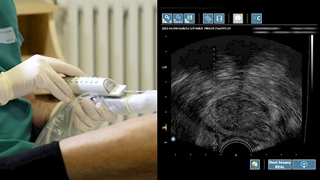
The transperineal approach involves an ultrasound probe inserted into the patient’s rectum (patients can be under general or local anesthesia). Motorized ultrasound transducers then capture the image of the prostate and display the ultrasound image onto the medical device used by urologists. Using MRI-ultrasound fusion, the previous MRI is loaded onto the system and fused with the ultrasound image, displaying a 3D view of the prostate gland.
“The TP approach evolved in efforts to maximize diagnostic accuracy and minimize morbidity. (Schmeusser, 2022)”
Once MRI fusion is achieved, physicians can now plan out their biopsy core targeting. The standard amount of prostate tissue samples is anywhere from 10-15 cores, depending on the stage of cancer and several other factors. The physician uses a biopsy needle and, through a prostate biopsy grid or freehand, inserts it into the patient via the perineum. The transperineal approach avoids piercing the rectal wall, which means it is often safer and less painful than the transrectal method of prostate biopsy.
After all cores are extracted, the samples are prepared to be sent to pathology for analysis. The patient is cleaned up and given instructions on post-biopsy follow up care. If future intervention is required, the patient may undergo focal therapy treatment to destroy cancerous cells.
When is a Transperineal Prostate Biopsy Needed?
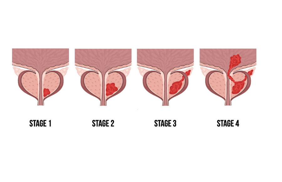
Above: Various stages of prostate cancer, from localized to advanced.
The transperineal biopsy is typically needed after a patient shows signs of having a risk of localized or advanced prostate cancer. One of the primary indicators of the presence of prostate cancer is elevated prostate-specific antigens (PSA) in the blood. Older men have routine PSA blood tests, and patients showing elevated PSA levels are often required to undergo a prostate biopsy.
However, not all urologists and practices implement the transperineal approach. The transperineal prostate biopsy typically requires advanced MRI-ultrasound fusion devices that are expensive, which means not all urology practices and hospitals can afford them.
Nonetheless, patients with suspected prostate cancer preparing for their biopsy can ask their doctor about the Koelis Trinity MRI fusion system. This platform uses Organ-Based Tracking® to track the prostate during the procedure for precision accuracy and 3D visualization. With the Trinity system, physicians can pinpoint suspected lesions with confidence and provide patients with the highest level of care.
Transperineal Fusion Prostate Biopsy
Not all transperineal prostate biopsies are created equal. As you learned, early versions of this method involved incisions through the perineum and were susceptible to heavy bleeding and infection. But today, ultrasound fusion technology allows for a minimally invasive biopsy that offers several advantages over other common biopsy methods.
With a transperineal fusion biopsy, MRI scans of the prostate are fused with the real-time ultrasound image of the prostate gland. This innovative fusion technology allows the physician to visualize the prostate in 3D and precisely biopsy suspected areas of the prostate containing cancer. The fusion biopsy also enables physicians to avoid unnecessary biopsies of other areas, which other methods may not have the capability to do.
Transrectal vs Transperineal Prostate Biopsy

We’ve talked a lot about the transperineal prostate biopsy and briefly mentioned the transrectal approach, but we haven’t touched on the differences between transrectal vs transperineal prostate biopsy directly. Let’s explore the main differences, benefits, and drawbacks to both approaches.
Transrectal Biopsy
The transrectal biopsy is another method of screening for prostate cancer that involves biopsy needles piercing through the rectum to obtain tissue samples in the same way the transperineal approach does. However, the biopsy needle enters the prostate gland at a different angle compared to the transperineal approach and may only need to be inserted as few as twice (but sometimes up to 24 times).
Nearly all patients undergoing a transrectal biopsy are prescribed antibiotics due to the risk of infection. Since the biopsy needle must pass through the rectal wall, each biopsy sample taken introduces the risk of rectal bacteria being introduced into the blood stream. Additionally, one study notes that patients claimed the transrectal biopsy was “the worst part of their prostate cancer journey”.⁴
Transperineal Prostate Biopsy Recovery Time

Recovery time for a transperineal prostate biopsy is relatively quick, with patients resuming normal activity within one to two days. According to a 2023 study, the risk of infection with the transperineal approach was 0.6% among 665 men undergoing transperineal prostate biopsy².
The transrectal biopsy recovery time is similar to its transperineal counterpart, however soreness or discomfort near the rectum is more common. The physician will recommend patients to avoid strenuous exercise for three to four days and may caution patients that blood in the urine and semen are common.
Final Thoughts
The transperineal prostate biopsy is an innovative approach to prostate cancer screening. What once involved painful incisions to the perineum has evolved into a technologically advanced biopsy method with specialized medical equipment and MRI fusion technology.
Regardless of the biopsy approach, prostate cancer screening has never been more effective. However, physicians using the transperineal prostate biopsy will find that this minimally invasive procedure is more effective at cancer targeting and detection and results in lower patient discomfort and complications².
Ask your urologist about KOELIS, or find a certified provider in our network with our KOELIS Locator.
Sources & References
1 – Schmeusser B, Levin B, Lama D, Sidana A. Hundred years of transperineal prostate biopsy. Ther Adv Urol. 2022 May 21;14:17562872221100590. doi: 10.1177/17562872221100590. PMID: 35620643; PMCID: PMC9128053.
2 – Grummet J, Pepdjonovic L, Huang S, Anderson E, Hadaschik B. Transperineal vs. transrectal biopsy in MRI targeting. Transl Androl Urol. 2017 Jun;6(3):368-375. doi: 10.21037/tau.2017.03.58. PMID: 28725578; PMCID: PMC5503965.
3 – Boesen L, Nørgaard N, Bisbjerg R, Al-Hamadani MMN, Sjölin CS, Løgager V. Office-based Magnetic Resonance Imaging-guided Transperineal Prostate Biopsy Without Antibiotic Prophylaxis: A Real-world Clinical Utility Study. Eur Urol Open Sci. 2023 Dec 23;59:71-77. doi: 10.1016/j.euros.2023.12.002. PMID: 38298768; PMCID: PMC10829603.
4 – Covin B, Roumiguié M, Quintyn-Ranty ML, Graff P, Khalifa J, Aziza R, Ploussard G, Portalez D, Malavaud B. Refining the risk-stratification of transrectal biopsy-detected prostate cancer by elastic fusion registration transperineal biopsies. World J Urol. 2019 Feb;37(2):269-275. doi: 10.1007/s00345-018-2459-4. Epub 2018 Aug 25. PMID: 30145777.
5 – https://www.cancer.org/cancer/types/prostate-cancer/detection-diagnosis-staging/tests.html