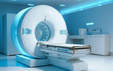
According to the National Cancer Institute, an MRI-guided biopsy is a procedure that uses an MRI scan to find lesions in prostate to guide the removal of a sample from the right area with a needle. Since 2022, international authorities recommend to use MRI/US system for a better diagnosis of prostate cancer. Zoom on the benefits of this procedure for urologists, but also and especially, patients.
The classical diagnosis of prostate cancer
In the diagnosis of most cancers, suspicious lesions that appear on endoscopic, CT-guided or ultrasound-guided observations are sampled. For prostate cancer, it is different. Guidelines recommend to perform systemic biopsies on patients who have a PSA level exceeding 4.0 ng/ml. These biopsies are performed under ultrasound guidance. To increase the probabilities to reach the lesions, 10 to 12 samples are collected from the all prostate.
This procedure shows limits:
✔️ First, for patients with early prostate cancer, the lesion is not visible under ultrasound guidance. This means that some patients are under diagnosis without any additional tests.
✔️ Second, systematics biopsies can miss, under or over evaluate the size of the lesion. Then the diagnosis is altered, and the treatment also.
The imprecise detection and grading of conventional systematic lead to over and under diagnosis of clinically significant prostate cancer. The treatment is also impacted. To avoid this, some physicians choose to do more biopsies to be sure, but this causes physical and mental anguish to the patient. Other choose to use MRI for a better diagnosis.
MRI fusion biopsy: which features are essential?
To make the diagnosis reliable, ultrasound images can be fused with MRI scans. It allows a better location of the lesion, thereby overcoming the disadvantages of conventional diagnosis. But to be efficient, some features are essential:
✔️ Elastic fusion: even if urologists use to do cognitive fusion, software elastic fusion gives them more precision and makes the diagnosis more reliable. MRI and ultrasound scans will automatically fuse to highlight the prostate and lesion location, increasing the cancer detection.
✔️ 3D representation of the prostate: the 3D mapping allows the physician to precisely locate the lesions and guide the needle.
✔️ Realtime location of the lesion: thanks to the scanning of prostate at each biopsy, and the fusion of images, physicians can locate the lesion during the all procedure, compensating for prostate movements.
✔️ Compensation of movement and prostate deformation: patient often move during the exam, due to his position, and the prostate can be distorted by the probe… So, it is sometimes difficult to know if the biopsies are realized into the lesion area. Fusion system should show adapt images to these movements.
All these features give urologists all the keys to be more efficient and trust their diagnosis to offer patient comfort and personalized treatment.
Koelis Trinity®: the right choice?
The Koelis Trinity® system meets all the expectations to help doctors in their daily work. Thanks to higher precision, physicians gain time and accuracy in their practices. Patient does not need to stay several days to hospital, they can treat more people.
The patented OBT™ (Organ-based tracking) technology, combined with 3D probes, consists on realizing new images at each biopsies to compensate prostate deformations. How does it work ? By matching 3D ultrasound image with the original reference value. Then, when the biopsy is performed, OBT™ shows the exact location of the cores and updates.
This device, and particularly OBT, allows urologists to be more precise with less samples, to have a better detection rate of prostate cancer and to perform less prostatectomies thanks to a better diagnosis, so a better treatment.
Thanks to the needle guidance, Koelis Trinity® offers physicians several options to perform biopsies and needle-guided treatments: transrectal or transperineal approaches, local or general anesthesia… Thus, patients can choose, in collaboration with their doctors, what fit best their expectations and needs.
To conclude, MRI targeted biopsies are a good option to a better detection of prostate cancer and for a higher accuracy in the diagnosis. This avoids under and over diagnosis and allow physicians to provide the best treatment for their patients. Among the options, active surveillance is often a great alternative and improve patient comfort while observing the progression of the cancer. Focal treatment is also an option that is more studied by physicians to preserve patient’s quality of life by treating only the target while preserving healthy tissues.
Sources :
Dr Tsutomu Tamada & Dr Yoshiyuki Miyaji user voices
https://ec.europa.eu/commission/presscorner/detail/fr/ip_22_5562
https://www.cancer.org/cancer/types/prostate-cancer/detection-diagnosis-staging/how-diagnosed.html
 United States

