English
 German
 Spanish
 Italian
 Japanese
 United states
What is the Gleason Score?

History of the Gleason Score
The Gleason Score is a system used to assess the aggressiveness of prostate cancer by examining prostate tissue under a microscope. Developed by American pathologist Donald Gleason in the 1960s, this scoring method classifies prostate tissue based on its deviation from normal patterns as it becomes cancerous.
Over time, numerous clinical studies have validated the Gleason Score as a reliable predictor of prostate cancer-specific survival.
Gleason Score Grading System
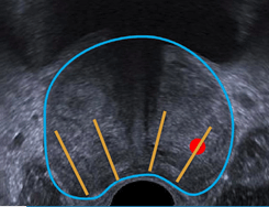
Pathologists assign Gleason scores ranging from 1 to 5 to individual biopsy samples or prostate specimens after surgery (see Figure 1). A score of 1 indicates tissue that closely resembles normal prostate tissue, while a score of 5 signifies highly abnormal, aggressive cancer cells.
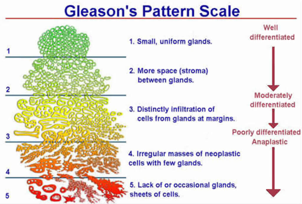
Figure 1: Schematic diagram of the Gleason score for tissue classification
The standard method for reporting the Gleason Score involves adding the two most prevalent patterns observed in the tissue sample.
For example, a Gleason Score of 3+3 indicates that pattern 3 is predominant, while a score of 3+4 means pattern 3 is dominant, but some pattern 4 is also present. If pattern 4 is more prevalent than pattern 3, the score would be 4+3. The total Gleason Score is the sum of these two numbers, ranging from 6 (3+3) to 10 (5+5).
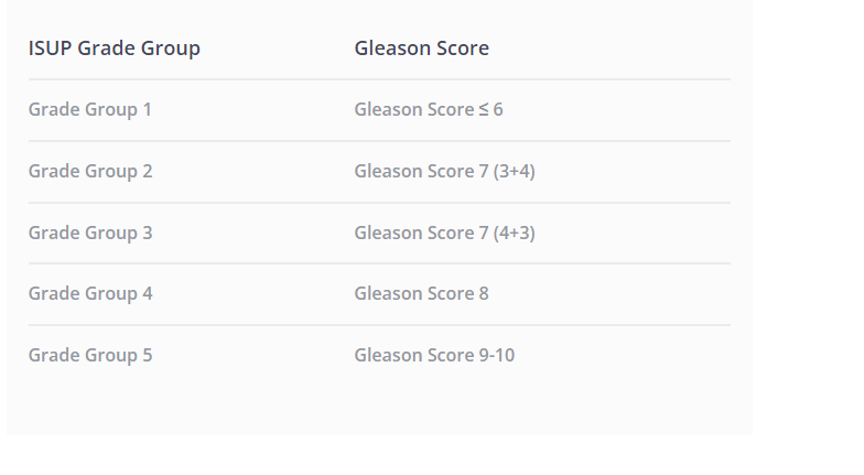
The ISUP Grade Group Classification
In November 2014, the International Society of Urological Pathology (ISUP) introduced the ISUP Grade Group classification to provide a clearer and more patient-friendly system. This classification aligns with the Gleason Score and is divided into five groups (see below).
Why Is the Gleason Score Important?
The Gleason Score is critically important because it serves as the gold standard for evaluating the aggressiveness of prostate cancer, guiding treatment decisions, and predicting patient outcomes. Here’s why it matters:
1. Determining Cancer Aggressiveness
- The Gleason Score helps classify how abnormal the prostate cancer cells are compared to normal cells.
- Lower scores (≤6) indicate slow-growing cancer, while higher scores (8-10) suggest aggressive cancer with a higher likelihood of spreading.
2. Guiding Treatment Decisions
- Patients with a low Gleason Score (6) may be candidates for active surveillance rather than immediate treatment.
- Higher scores often lead to more aggressive treatments, such as radiation therapy, hormone therapy, or surgery.
3. Predicing Patient Outcomes
- Studies have shown that the Gleason Score strongly correlates with survival rates.
- A higher score means a greater risk of cancer recurrence and metastasis.
4. Standardized and Globally Recognized
- The International Society of Urological Pathology (ISUP) introduced Grade Groups based on the Gleason Score to make risk classification clearer for patients and doctors.
- The Gleason Score remains a widely accepted tool used in conjunction with PSA levels, imaging, and biopsy results.
Drawbacks of the Existing Diagnostic Methods
Traditional 2D ultrasound-guided biopsy has several limitations that can impact the accuracy of prostate cancer diagnosis. The key drawbacks include:
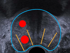
a) In some cases, the biopsy completely misses the cancerous area, leading to a false-negative result.
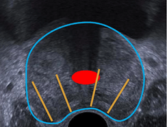
b) The biopsy may capture only a small portion of a clinically significant tumor which can underestimate the aggressiveness of the cancer.
c) In some cases, the biopsy targets a non-clinically significant tumor, leading to overdiagnosis and overtreatment that may cause unnecessary side effects.
The advent of MRI facilitated the development of fusion systems that integrate MRI with ultrasound and improved lesion targeting and biopsy accuracy. However, conventional MRI/ultrasound fusion systems primarily utilize rigid fusion technology which does not account for prostate deformation or patient movement.
As a result, the MRI-identified target may be misaligned, leading to potential discrepancies in biopsy sampling and a false assumption of precision in targeting suspected lesions.
How KOELIS® improves prostate cancer diagnosis1
The KOELIS Trinity® Prostate Fusion Biopsy System integrates cutting-edge technologies to enhance diagnostic accuracy2, biopsy traceability, and treatment planning.
3D Prostate Mapping for Enhanced Diagnosis
KOELIS Trinity® creates a personalized 3D map of the prostate for each patient, incorporating:
- The prostate contour
- Suspicious MRI-identified areas
- Biopsy core locations
This advanced mapping provides a comprehensive view to assist clinicians in precise targeting and treatment decision-making.
Patented OBT Fusion® Technology for Unmatched Accuracy
The OBT Fusion® technology provides millimetric precision2 by compensating for prostate deformations and patient movements during biopsy. Our OBT technology significantly enhances the reliability of 3D mapping and biopsy localization.
Seamless MRI/Ultrasound Elastic Fusion for Precision Biopsy
By combining MRI/US elastic fusion with real-time 3D ultrasound, the KOELIS Trinity® system enables highly accurate and targeted biopsies through either a transperineal or transrectal approach. This precision provides:
- A more accurate assessment of prostate cancer stage
- Patient-specific treatment planning tailored to individual risk levels
Top Cancer Centers Trust KOELIS


Learn More
Sources and Links
2 https://koelis.com/academy/precision-matters-in-mr-imaging-targeted-prostate-biopsies/