
 English
 German
 Spanish
 Italian
 Japanese
 United states
Prostate cancer is the second most common type of cancer among men in the United States, and nearly 1 in 8 men will receive a prostate cancer diagnosis in their lifetimes. However, our ability to screen and detect prostate cancer has never been stronger.
With a prostate biopsy, urologists are able to sample specific regions of the prostate suspected of containing cancer, often using MRI-ultrasound guidance for enhanced accuracy. Samples taken by the urologist are sent to pathology to analyze the presence of cancerous tissue within the prostate.
With today’s advanced tools and technological innovations, a prostate biopsy is a safe and effective way to screen for prostate cancer. Let’s find out the truth about prostate biopsy and talk about the procedure, its painfulness, and the accuracy of prostate biopsies.
How is a Prostate Biopsy Done?
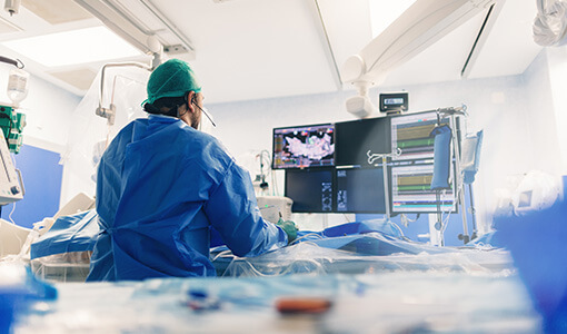
At KOELIS, our Trinity® system uses MRI-ultrasound fusion technology for precise accuracy during a prostate biopsy. Here is the full prostate biopsy procedure description using the KOELIS Trinity® fusion biopsy system.
Prostate Biopsy with KOELIS Trinity®
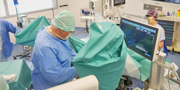
The patient is positioned in either the lithotomy or left lateral decubitus position depending on whether a transrectal or transperineal approach is used. The area is prepped and draped using standard sterile technique. Local or general anesthesia is then administered.
The ultrasound probe is attached to the MRI-ultrasound system via a mechanical stepper that starts the automated tracking of prostate motion. An ultrasound scan of the prostate is then acquired to capture the live gland.
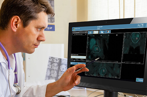
Multiparametric MRI data from a prior diagnostic exam are loaded into the Trinity® platform. Regions of interest (ROIs) identified on MRI are fused with the real-time ultrasound scan using KOELIS’ proprietary Organ-Based Tracking® (OBT) technology. This fusion accounts for prostate deformation, motion, and patient positioning for a highly accurate overlay of MRI targets on live ultrasound images.
Once fusion is confirmed, the physician selects target lesions from the MRI overlay and defines the number and location of cores to be extracted. All core trajectories are displayed in real-time 3D within the prostate volume.
Biopsy sampling is performed under continuous ultrasound guidance. The biopsy needle is aligned with the probe guide (transrectal) or introduced through the perineal grid or freehand (transperineal). The KOELIS system tracks the needle path and updates the 3D model dynamically while recording the spatial position of each core.
Once all planned cores have been obtained, the probe is withdrawn, the area is cleaned, and the patient is monitored briefly for any immediate complications. Patients are discharged with post-procedure care instructions. Transrectal cases may involve short-term antibiotics. Transperineal cases typically do not require catheterization or prophylactic antibiotics. Most patients resume normal activity within 24–48 hours.
The KOELIS biopsy map is saved for use in longitudinal care such as follow-up biopsies, active surveillance, or focal therapy planning.
Is a Prostate Biopsy Painful?

A prostate biopsy is a minimally invasive procedure that doesn’t involve large incisions and can be done in an outpatient facility. Recovery times are typically fast and patients are often back to resuming normal daily activities within a few days.
Whether the procedure is done transrectally or via the transperineal route, a prostate biopsy is generally uncomfortable but not very painful. Patients feel a quick pinch or pressure each time the biopsy needle enters the prostate, resulting in mild to moderate discomfort.
When done transperineally, the prostate biopsy is often less painful since it’s done with general anesthesia or deep sedation. Patients will likely feel soreness around the perineum for a few days following the procedure. Men often describe the sound of the spring-loaded needle more startling than painful.
However, it’s important to understand there are several factors that can influence whether or not the prostate biopsy is painful. The transrectal method tends to be more uncomfortable for patients since the needle passes through the rectal lining. Additionally, the number of samples taken and patient anxiety are factors.
How Long is a Prostate Biopsy?
A transrectal prostate biopsy is the fastest type of biopsy and is often completed in 10-20 minutes. However, when using MRI-ultrasound fusion technology, the procedure itself may take 45 minutes or longer, and time spent at the clinic or hospital may be closer to 1-2 hours to account for preparation and post-procedure observation.
Who Needs a Prostate Biopsy?
Men suspected of having prostate cancer undergo the prostate biopsy to formally test for the presence of cancer. This is often first detected after a prostate-specific antigen (PSA) blood test¹, although high PSA levels can also be an indicator of benign prostatic hyperplasia (BPH) or prostatitis.
Additionally, men undergoing treatment for prostate cancer may have repeat biopsies to rule out the possibility that cancer has spread or to confirm that the prostate cancer is under control or in remission. Specifically, men who were treated with focal therapy or put on active surveillance and show high PSA levels may warrant a repeat biopsy.
“Men suspected of having prostate cancer undergo the prostate biopsy to formally test for the presence of cancer. This is often first detected after a prostate-specific antigen (PSA) blood test.”
How Accurate is a Prostate Biopsy?
Prostate biopsies are generally fairly accurate, although the method of biopsy and medical equipment are factors. A 2013 study notes that prostate biopsy is accepted as the best diagnostic technique to detect prostate cancer², but specifically the transperineal route with MRI-ultrasound fusion technology showed to be more effective at cancer detection³.
The number of cores extracted is also a major factor in the accuracy of the prostate biopsy. More cores extracted means a higher chance of finding cancer. Additionally, a pre-biopsy MRI improves cancer detection and is required for MRI-ultrasound fusion biopsies.
False negatives and repeat biopsies remain an issue, as a negative biopsy does not always mean no cancer. The cancer may be clinically insignificant, low-grade, or localized and can be missed with a prostate biopsy⁴. If PSA levels remain high, repeat biopsy may be necessary.
The Bottom Line
This article uncovered the truth about prostate biopsy, discussing the procedure, length of the prostate biopsy, the level of pain experienced, and the accuracy of biopsies. Whether you’re a patient, loved one of someone with prostate cancer, or a medical professional, understanding the truth about prostate biopsy is essential.
Ask your urologist about KOELIS, or find a certified provider in our network with our KOELIS Locator.
Sources & References
1 – Prostate-Specific Antigen (PSA) Test: Purpose & Results. Cleveland Clinic. https://my.clevelandclinic.org/health/diagnostics/24615-psa-test
2 – Serefoglu EC, Altinova S, Ugras NS, Akincioglu E, Asil E, Balbay MD. How reliable is 12-core prostate biopsy procedure in the detection of prostate cancer? Can Urol Assoc J. 2013 May-Jun;7(5-6):E293-8. doi: 10.5489/cuaj.11224. Epub 2013 May 13. PMID: 22398204; PMCID: PMC3668408.
3 – Cornud F, Roumiguié M, Barry de Longchamps N, Ploussard G, Bruguière E, Portalez D, Malavaud B. Precision Matters in MR Imaging-targeted Prostate Biopsies: Evidence from a Prospective Study of Cognitive and Elastic Fusion Registration Transrectal Biopsies. Radiology. 2018 May;287(2):534-542. doi: 10.1148/radiol.2017162916. Epub 2018 Jan 22. PMID: 29361246.
4 – M. Klingebiel, C. Arsov, T. Ullrich, et al. Reasons for missing clinically significant prostate cancer by targeted magnetic resonance imaging/ultrasound fusion-guided biopsy. European Journal of Radiology. Volume 137. 2021. 109587. ISSN 0720-048X. https://doi.org/10.1016/j.ejrad.2021.109587.