
Welcome to KOELIS®
Please choose a country or region.
-

US -

Global

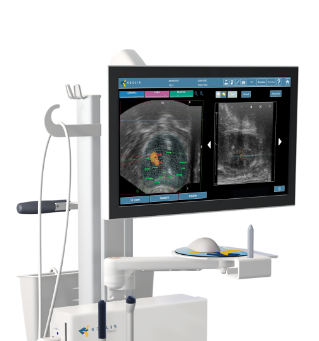
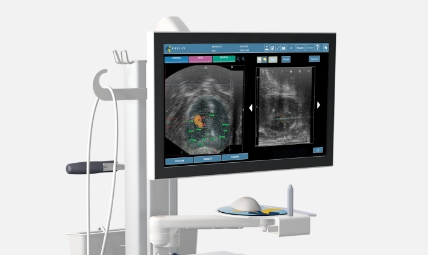
KOELIS Trinity® is a comprehensive solution for urologic patient management, from screening to diagnosis and treatment. KOELIS Trinity® fusion biopsy system integrates with KOELIS’ ProMap Lite™ software that allows radiologists to conduct real-time analysis of MRI and mpMRI of the prostate, report those findings and easily prepare that data for the urologist performing prostate fusion biopsy.
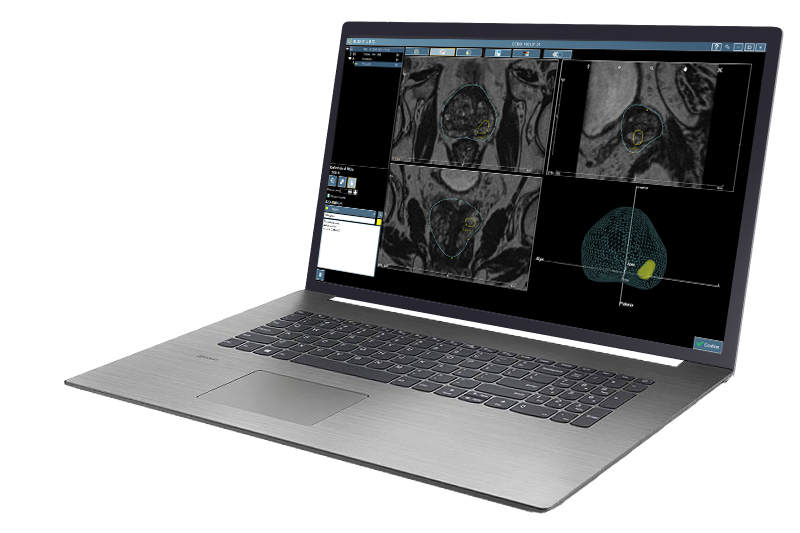
For planning and evaluating the most accurate and personalized prostate cancer strategy, measurement is a key factor. KOELIS ProMap Lite™, an intuitive and easy-to-use interface, allows you to easily contour the prostate and target lesions as well as to define their volume measurements for millimetric precision. Mark lesions and risk zones effortlessly directly on the imported multiparametric images as well as retrieving previous information for assessment.
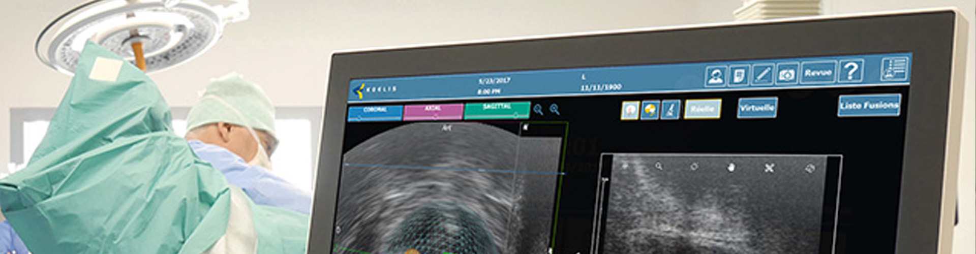
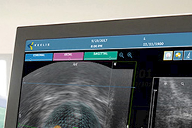
ProMap Lite™ allows for the fusion of two imaging modalities offering physiologic and functional information combined, while matching anatomical points, so targets are displayed with the highest accuracy. Clinicians can transfer contours from one image to another, as well as configure transparency to support diagnosis’ needs.
Measurement of Standardized Uptake Value (SUV) can be marked on PET images for assessing the activity in a given focus and potentially improve the accuracy and consistency of lesion identification. Clinicians use this feature in ProMap Lite™ to define standards for lesion staging, localize tumors with accuracy, and define contours and margins with total confidence.
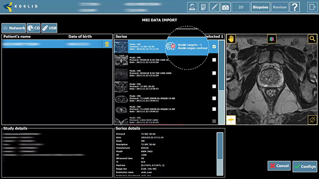
Urology, radiology and nuclear medicine can now work more efficiently for better patient selection and treatment planning. ProMap Lite™ makes it possible to immediately send and receive a patient’s images, KOELIS 3D cartography and other valuable information from the PACS, as well as to export it into DICOM Surface Segmentation format with a simple gesture.
The connected system facilitates improved and more consistent communication between specialists, even in remote sites, that promotes better prostate cancer detection and management. All the updated information can be fast stored and shared to bring quality control to future interventions’ planning or patient monitoring.
© 2024 KOELIS®. All rights reserved. KOELIS Trinity® is a registered trademark of KOELIS® in certain countries.

Request a live demo today