

 English
 German
 Spanish
 Italian
 Japanese
 United states
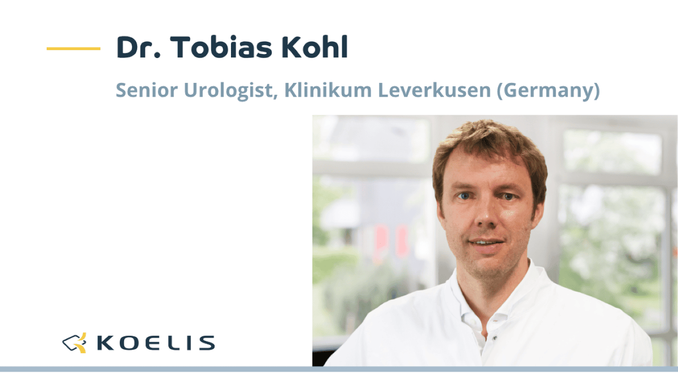
Dr. Tobias Kohl, Senior Urologist at Klinikum Leverkusen (Germany), has been performing fusion biopsies for more than ten years. His team performs around 800 procedures each year using the transperineal approach.
Improving image preparation
Before each biopsy session, MRI images need to be prepared and the prostate contoured to ensure accurate MRI/ultrasound fusion. While this step is essential, it can also be time-consuming and its quality often depends on the operator’s level of experience.
After introducing ProMap® Smart into his workflow, KOELIS’ AI-powered automatic segmentation solution, Dr. Kohl wanted to evaluate how AI could support this part of the workflow.
His first impression was straightforward.
“The accuracy of the automatically generated contours was remarkably high, and the speed was striking. It immediately felt like a tool that was ready for routine clinical use.”
Supporting everyday clinical practice
Beyond the time saved, Dr. Kohl highlights the impact on his whole team. Less experienced users can achieve a consistent level of contouring more quickly, helping reduce variability between operators.
“ProMap Smart has accelerated the workflow for every member of our team. The gap between beginners and experts has become much smaller, directly improving the consistency and reproducibility of our procedures.”
He also notes that integrating the solution into daily practice required no change to the existing workflow.
“You simply open the prostate images, whether TRUS or MRI, and the contours are already there.”
As AI continues to be integrated into prostate fusion procedures, clinicians remain in control while benefiting from tools designed to simplify image preparation and support a more consistent workflow.