English
 German
 Spanish
 Italian
 Japanese
 United states
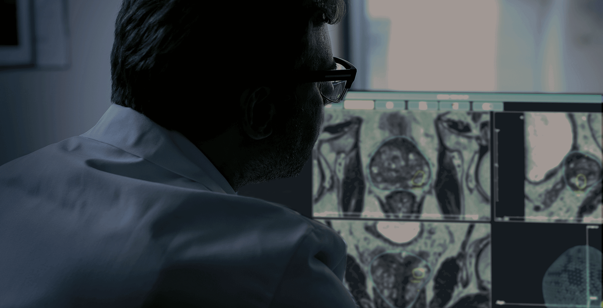
Prostate biopsy is a medical examination that consists of collecting small prostate samples to analyze them to confirm whether an anomaly is benign or malignant. This step is essential to confirm a prostate cancer diagnosis.
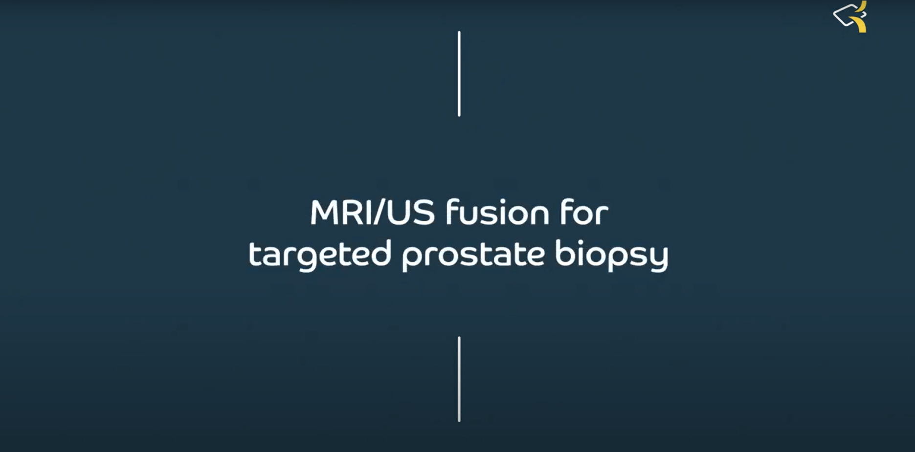
Year after year, approaches and methods have evolved to be more effective and to improve cancer detection and patient comfort. Ones of them are targeted biopsies based on MRI/US fusion. Let’s talk about it!
Prostate biopsies: where everything has began
For a better understanding of targeted biopsies, it is necessary to look back to the birth of prostate biopsies. There are born in the 80s in the USA. First, they were performed by transperineal approach. The famous urologist Dr Hodge has the idea to use the endocavitary probe to use the transrectal approach. This has triggered a lot of debates over whether ultrasound images could detect prostate cancer. Then, Dr Hodge has had the idea to create a prostate biopsy map. That means to take 6 cores from the right lobe and 6 from the left.
In the 90s, the debate has been on the number of cores to collect. Some researchers have concluded that 12 cores for transrectal approach enable the prostate to be sampled homogeneously to diagnose a cancer smaller than 1cm3. It does not work for patients with large prostates and with a rising PSA. The number of samples was about 20 and 30, and the conclusion was often prostate recession or adenomectomies.
Some physicians have decided to use transperineal approach with a grill guide to collect cores. This also end to a problem: there were a saturation of biopsies (up to 50 biopsies by patient) with side effects: acute urine retention.
What is MRI-Targeted prostate biopsy?
With progresses in the health industry, some radiologists have had the idea to use some techniques developed in other specialties (cardiology mainly) for prostate biopsy. This is how MRI images have been used in urology.
Two major branches have been created:
- – Biopsy directly in the MRI ring
- – Use of echography and develop a software to merge MRI and US images, and then perform the prostate biopsies.
Today, the second branch is the most used.
Which technologies exist for targeted biopsies?
The images fusions methods have known major developments. Today, there is two images fusion methods:
- – Rigid fusion: points are taken from the MRI (APEX, base et posterior part of the prostate), then an echography acquisition is performed. With this kind of method, we are expecting an accuracy in the cm range, with a very important human factor for selecting the same points in the MRI and ultrasound images. It requires a considerable expertise, often resulting in significant inter-operator variability in fusion accuracy results.
- – Elastic fusion: there is no minimum number of points, but it generates mesh in MRI and US images. We’ll have two points clouds that will match these point clouds thanks to technologies and software. The result is a realignment of the organ. These are today the most developed and advanced technologies.
Expected results of targeted prostate biopsies
Results depend on the MRI quality. A well-done MRI with a PiRADS 5 will give better results than a less well-done MRI with a PiRADS 3.
Experience at Pitié Salpétrière (published study)
Using Koelis Technology, based on elastic fusion with OBT technology
- – If a patient is PiRADS 3, the probability of having a positive core in the target is 20% ; if he has a PiRADS 5, the probability is 95% ; if PiRADS 4, it’s 60%.
- – It changes the dialog with the patient on the expected results of biopsies.
- – 10 years ago, a patient who came to the clinic had a 40% probability of having a positive biopsy. Today, with MRI images and targeted prostate biopsies, there is almost 80% of chance to have at least one positive core.
To conclude, it is necessary to follow the recommendations, that evolves with time. Today, the standard is making 12 systematic cores and targeted cores.
Sources :
- Podcast de Pr Pierre Mozer (https://open.spotify.com/episode/4Q8Z7iWYHu0CpG2A1Uqorx)
- https://pubmed.ncbi.nlm.nih.gov/25663102/